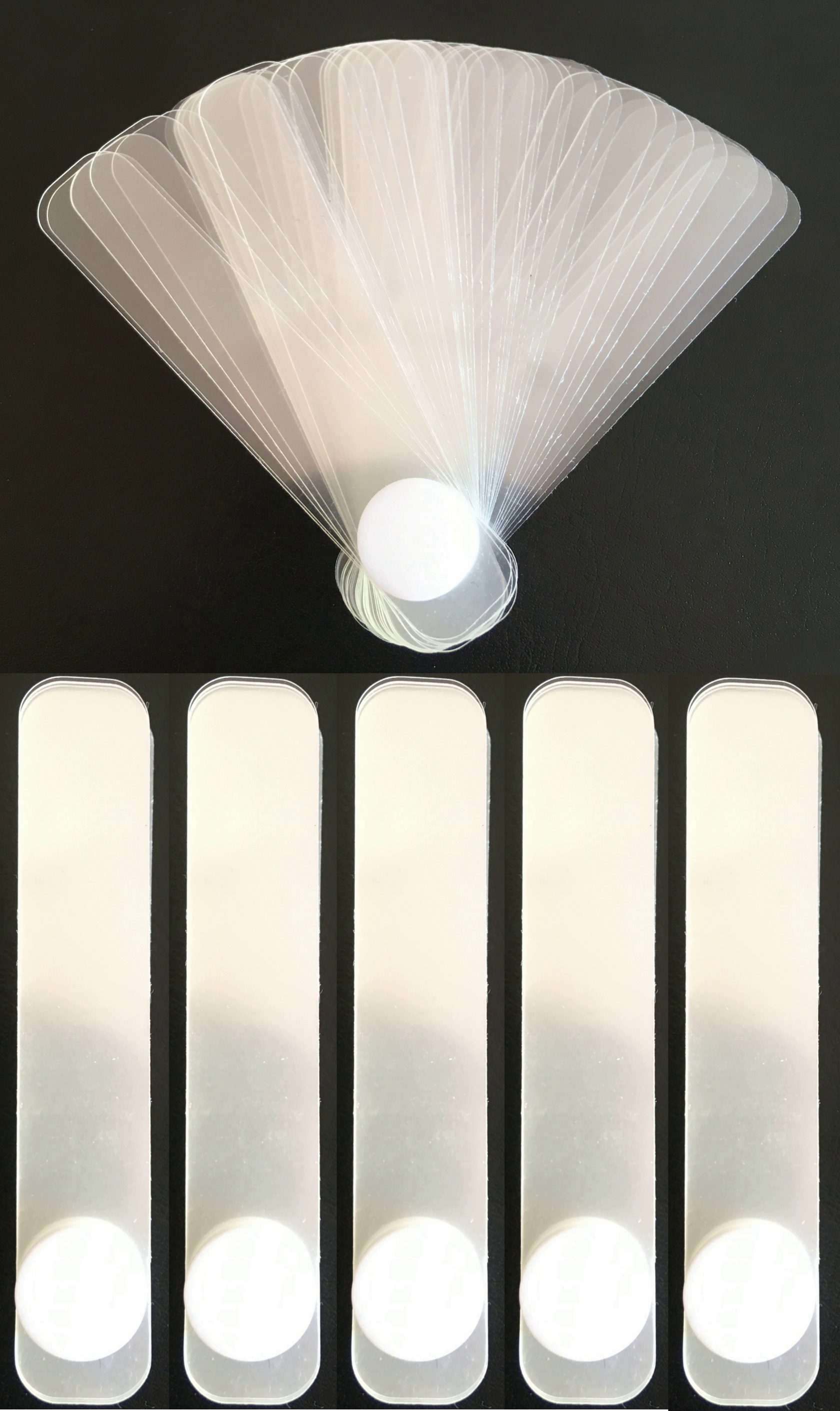
We produce in Russia and we ship all over the world
Leaf gauges and Lucia Jig
Manufacture and sale of instruments that are used by most dentists around the world to identify and register the correct position of the mandible, for stress testing and selective grinding
To order 
Usage guide of leaf gauges and Lucia Jig

In 1973, James H. Long proposed the use of an anterior deprogrammer in the form of a fan of sheets as a device to align and center the mandible and normalize occlusion. Over the course of several decades, numerous method adjustments have been made and as a result, several protocols have been developed for the use of the leaf gauge by several dental schools in the United States and Europe.
Until 2017, leaf gauge production was carried out only in the USA and Canada.
In 2017, the production of leaf gauges and Lucia jigs was launched in Russia, and a utility model patent was obtained.
Until 2017, leaf gauge production was carried out only in the USA and Canada.
In 2017, the production of leaf gauges and Lucia jigs was launched in Russia, and a utility model patent was obtained.

In order to find the centric relation (in the case of patients without a pronounced anatomical pathology of the joint), it is necessary to remove overstrain, excessive tone of the lateral pterygoid muscle on both sides, as a result of which the positioning of the lower jaw due to the articular elements in the position of the central ratio will occur. To eliminate the overstrain of the lateral pterygoid muscle, it is necessary to eliminate the proprioceptive sensitivity of the teeth and remove the effect of super contacts. To eliminate the proprioceptive sensitivity of the teeth, it is necessary to separate them using anterior deprogrammers - a leaf gauge and a jig Lucia
The positioning a leaf gauge or jig in the area of the anterior teeth, the masticatory group of teeth is dissociated, as a result of which there is a lack of proprioceptive sensitivity in the area of these teeth, then the relaxation of the lateral pterygoid muscle occurs, which, until the moment of relaxation, positioned the location of the lower jaw in the position of the usual occlusion. Due to the relaxation of the lateral pterygoid muscle, the mandible is positioned in the place of the central relationship due to the fact that the lower jaw at this moment has only three points of support - a leaf gauge or jig Lucia in the area of the anterior teeth and elements of the temporomandibular joint dysfunction. In the absence of displacement due to the lateral pterygoid muscle, the elements of the temporomandibular joint dysfunctions are positioned in a physiologically correct position (if there are no pathological changes in the structure of the joint structures).
The positioning a leaf gauge or jig in the area of the anterior teeth, the masticatory group of teeth is dissociated, as a result of which there is a lack of proprioceptive sensitivity in the area of these teeth, then the relaxation of the lateral pterygoid muscle occurs, which, until the moment of relaxation, positioned the location of the lower jaw in the position of the usual occlusion. Due to the relaxation of the lateral pterygoid muscle, the mandible is positioned in the place of the central relationship due to the fact that the lower jaw at this moment has only three points of support - a leaf gauge or jig Lucia in the area of the anterior teeth and elements of the temporomandibular joint dysfunction. In the absence of displacement due to the lateral pterygoid muscle, the elements of the temporomandibular joint dysfunctions are positioned in a physiologically correct position (if there are no pathological changes in the structure of the joint structures).
Links to articles and publications
Russian - language:
- Центральное соотношение и центральная окклюзия: анализ взаимосвязей
- Процедура коррекции полноконтактной окклюзионной шины с передней направляющей
- Клинический протокол устранения окклюзионных балансирующих препятствий
- Сравнительная характеристика методов депрограммирования жевательных мышц
- Программированная координация работы жевательных мышц и положения нижней челюсти в лечении пациентов с функциональной патологией височно-нижнечелюстного сустава
English - language:
- A Clinical Protocol for the Removal of Balancing Interferences
- Leaf Gauge Refinements
- Three Options to Restore Patients in CR
- Evaluating 'At Risk' Occlusions
- Leaf Gauge and the Orthodontic Finish
If a person's centric relation and the usual occlusion coincide, then the work of joints, muscles, teeth becomes most effective, and all chewing loads are distributed most efficiently. This leads to a significant reduction in the risk of teeth chips, composite and ceramic restorations, removes the occlusal component of chronic gum inflammation. The coincidence of the centric relation and the usual occlusion also has a positive effect on increasing the stability of the work of the components of the temporomandibular joint.
The main role in the positioning of the lower jaw in space is played by the lateral pterygoid muscle. Unilateral contraction of this paired muscle leads to a displacement of the mandible in the opposite direction from which muscle contracted. Bilateral muscle contraction causes the lower jaw to move forward.
In the event of super contacts between the teeth due to proprioceptive sensitivity, reflex tension of the lateral pterygoid muscle occurs, which leads to a shift of the jaw away from early contact to eliminate overload and to bring multiple contacts to the position. With this displacement of the jaw, the joints gradually adapt to the new position.
Over time, a person has a lot of super contacts due to the violation of the position of the teeth, the appearance of fillings, crowns. The jaw is shifting more and more, the joints have to expand their adaptive capabilities. Unfortunately, adaptive capabilities have their limits, and at some point, many people experience a breakdown in compensation, as a result of which there is either tooth decay, or chronic inflammation of the gums, or problems with the temporomandibular joint and chewing muscles.
First, 5 pieces of gauge (i.e. 0.5 mm) are placed in the area between the teeth of the upper and lower jaw, we ask the patient to close the chewing teeth.
If at the same time the patient feels that the posterior teeth are closing, then add 5 more sheets, repeat the question and add leafs until the patient indicates that if there are leaf gauge between the front teeth, he does not feel the lateral teeth in contact.
After that, add another 5-6 leafs. Then we begin to move the lower jaw along the leaf gauge back and forth. At the moment of displacement of the lower jaw back, we ask the patient to try to compress the chewing teeth a little. After 5-6 minutes of such movements, the lateral pterygoid muscle, as a rule, relaxes, and the articular elements are positioned in their comfortable position - the position of the centric relation (if there are no anatomical changes in the joint area). Further, depending on the purpose of using the calibrator at the moment, either the registration of the obtained central ratio is carried out, or the search for super contacts.
The doctor begins to remove the leaf gauge one by one from the part that is located between the front teeth. To control whether the lateral teeth are closing, there is contact between them or not, a special occlusal copy paper 200 microns thick (to create a large contact patch) and paper 8 microns thick and different in color (to determine the super contact point area) are used. The described 200-micron occlusal paper is positioned between the posterior teeth, then the patient closes the teeth in a leaf gauge, repeats the back-and-forth movements. Then tries to close half the strength of the chewing teeth in the position of the lower jaw from behind. At this moment, the doctor is trying to get the paper between the teeth of the lateral sections. If he succeeds easily, then the doctor should remove one leaf from the part of the gauge sandwiched between the front teeth. Repeat movements. And so continue until the moment when it will be impossible to get the paper between the lateral teeth without opening them. At this point, the doctor positions a thin 8-micron paper between the posterior teeth. Asks to repeat the movements of the lower jaw. Continue to remove the leaf gauges until the 8 micron foil lingers on either side.
As a result, a thick contact spot is imprinted on the teeth due to 200-micron paper and a point supercontact on this spot due to 8-micron paper. This completes the search for a supercontact that provokes the displacement of the lower jaw.
If selective grinding is carried out, the doctor grinds this supercontact, after which it again positions the calibrator in the area of the anterior teeth, occlusal paper in the area of the posterior teeth, and checks for the presence of supercontact. If it disappeared after grinding, then the doctor removes another leaf of the gauge and continues to look for super contacts further according to the previously described method. Grinding is considered complete if 2-4 leaf gauges or more remain in the area of the front teeth (this parameter is very individual, since it depends on the patient's bite), and between the lateral teeth at the same moment there are multiple contacts on both sides of the jaw. The result is the adjustment of the position of the lower jaw in the usual occlusion to the position of the central ratio of the jaws.
Next - without a leaf gauge, check for canine and incisal protection and remove early contacts on the balancing side and on the working side on molars and premolars during lateral movements.
IMPORTANT - the use of the grinding technique with a leaf gauge in patients with distal occlusion is contraindicated, as it can lead to additional distalization of the mandible.
More products
Instructional Video
Below are videos on the use of leaf calibrators and jigs Lucia in the dentist's practice
Frequent Questions
No, leaf gauges can be used at any primary dental practice to perform a TMJ stress test to determine the condition of the joints. Also, as a quick way to identify super contacts.
Take an arbitrary number of leafs, position them between the anterior incisors and check if there are any contacts in the lateral sections when the lower jaw moves back and forth. If contacts are detected in the lateral sections, then it is necessary to add the number of sheets. Most often, a third of the gauges thickness is sufficient.
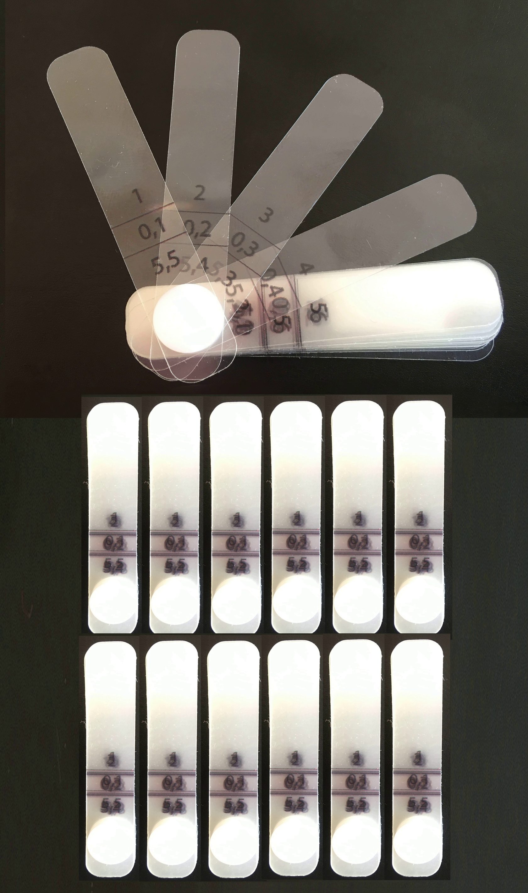
On the leaf gauge, each leaf is labeled. The upper digit is the number of the leaf. The lower one is the thickness of the total number of sheets taken starting from the first one.
Convenient for determining the degree of tooth separation and for monitoring during the search for early contact and selective grinding.
Convenient for determining the degree of tooth separation and for monitoring during the search for early contact and selective grinding.
Yes, leaf gauges are reusable.
For their processing, it is optimal to use autoclaving at 120 ° C at 1.1 atm for 45 minutes, or use an exposure in 6% hydrogen peroxide solution for 6 minutes
For their processing, it is optimal to use autoclaving at 120 ° C at 1.1 atm for 45 minutes, or use an exposure in 6% hydrogen peroxide solution for 6 minutes
Yes, gigs Lucia are reusable.
For their processing, it is optimal to use an exposure in 6% hydrogen peroxide solution for 6 minutes
For their processing, it is optimal to use an exposure in 6% hydrogen peroxide solution for 6 minutes
Delivery within the territory of the Russian Federation can be organized either by Russian Post or by the transport company CDEK
When ordering by post - the cost of postage is paid in advance. But you can pick up the parcel ONLY at the post office.
When ordering CDEK, the cost of delivery services is paid to the courier at the time of receipt of the order. Convenient if ordered to the clinic.
When ordering by post - the cost of postage is paid in advance. But you can pick up the parcel ONLY at the post office.
When ordering CDEK, the cost of delivery services is paid to the courier at the time of receipt of the order. Convenient if ordered to the clinic.
Yes, it is possible. But only by post office.
The optimal number is 2-3 leaf gauges per work shift, since they are used during the initial consultation and stress test on the temporomandibular joint dysfunction, to identify early contacts, selective grinding, quick correction of restorations by occlusion, grinding splints and splints. Therefore, it is enough for 1 doctor to order a set of 3 labeled leaf gauges as a starting point.
Jigs Lucia are used only at the stage of registration of the central ratio during diagnostics. As a rule, one set is enough for a long period of time.
Jigs Lucia are used only at the stage of registration of the central ratio during diagnostics. As a rule, one set is enough for a long period of time.
Typically, the lifespan of leaf gauge is about 1-1.5 years, after which there is a gradual deformation of the leafs.
Jigs Lucia can be used for about the same time. But, as practice has shown, they are often lost during cleaning the office after receiving patients.
Jigs Lucia can be used for about the same time. But, as practice has shown, they are often lost during cleaning the office after receiving patients.
Leaf gauges without numbers perform several functions - they are used for a load test on the TMJD, for deprogramming the muscle, for recording the centric relation.
Leaf gauges with numbers, in addition to the above, give accurate data on the degree of separation between the front teeth and this allows more accurate selective grinding and more convenient plaster cast into the articulator (if the doctor uses a leaf gauge for registration)
Leaf gauges with numbers, in addition to the above, give accurate data on the degree of separation between the front teeth and this allows more accurate selective grinding and more convenient plaster cast into the articulator (if the doctor uses a leaf gauge for registration)
About us in numbers
Results in 3 years
14
The number of countries
178
The number of cities
680
The number of customers
3350
The number of leaf gauges shipped
Our team
- Nikolay GranovskyProsthodontic, head of the Arche training club, co-founder of Byte-market, lecturerNikolay treats patients with complex musculo-articular dysfunctions. He founded the Arche training club in Arkhangelsk in 2018. Since 2017, he organized the production of leaf gauges and Lucia jigs in Russia.
- Yulia GranovskayaHead of the Byte Market. Co-founder of Byte Market. Handles orders and logistics.
Clinical psychologist, supervises the work of doctors with patients.Yulia is already known to many dentists, since she took over all the operational activities for the orders and delivery of sheet calibrators and Lucia jigs.
As a clinical psychologist, since 2005, she has been conducting psychological consultations, as well as supervision of medical appointments to improve the quality of work doctors.


